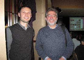
| H. Joseph Yost, Ph.D., research |
Research authored by Dr. Yost and his lab
Click the tab and a window will appear.
List of Publications via PubMed
(NIH National Library of Medicine)
List of Publications via PubMed
(NIH National Library of Medicine)
| ||||||||||||||||||||||||||||||||||||||||||||||||||||||||||||||||||||||||||||||||||||||||||||||||||||||||||||||||||||||||||||||||||||||||||||||||||||||||||||||||||||||||||||||||||||||||||||
| ||||||||||
2009 Conference abstract
HS Fine Structure and FGF Signaling Pathways Converge at Cilia: Does Cilia Function Have a Role in HME?
H. Joseph Yost, Ph.D.
Departments of Neurobiology & Anatomy, Pediatrics. University of Utah School of Medicine.
http://yost.genetics.utah.edu
Several peptide growth factors, including members of the FGF, TGFb, BMP and Wnt families, have been implicated in the
regulation of cartilage and bone growth. The functions of these factors are regulated in part by complex Heparan Sulfate
Proteoglycans (HSPGs) at the cell surface. Alterations in genes involved in synthesis of Heparan Sulfate (HS) chains, as
exemplified by mutations in EXT1 and EXT2, lead to Hereditary Multiple Exostoses (HME), perhaps by misregulating complex cell-
cell signaling pathways. Heparan sulfate (HS) is an unbranched chain of repetitive disaccharides, typically attached to the core
proteins of Heparan sulfate proteoglycans (HSPGs), which specifically bind peptide growth factors at the cell surface or secreted
extracellularly. HS chains contain sulfated domains termed the HS "fine structure" which are catalyzed by HS O-sulfotransferases
(OSTs).
Our working hypothesis is that there is a "Glycocode" embedded in the fine structure of HS, and that distinct members of the
HS 3-OST family contribute to distinct signatures within this code, which then regulate specific cell-cell signaling pathways. Using
gene knock-down screens in zebrafish, we have uncovered distinct roles for three members, 3-OST-5, 3-OST-6 and 3-OST-3Z
expressed in the same cell lineages. Strikingly, both 3-OST-5 and 3-OST-6 are required for distinct functions of cilia, which are
cell surface organelles found on most epithelia in vertebrates. Recently, cilia have been implicated as recipients of some cell-cell
signaling pathways, for example, Hedgehog signaling. However, little is known about cell-cell signaling pathways that control the
formation or function of cilia. We found that 3-OST-5 is required for normal cilia length, whereas 3-OST-6 controls cilia motility.
Knockdown of a third 3-OST family member has normal cilia function. In collaboration with Jeff Esko’s lab, we find that
knockdowns of each of these three 3-OST family members cause a similar reduction of a 3-O-sulfated disaccharide subunit in
the HS chains, so the differences in cellular phenotypes are not simply due to bulk changes in sulfation. Further analyses indicate
that 3-OST-5 and 3-OST-6 modulate distinct cell-cell signaling pathways in cilia formation or function, presumably by distinct
glycocodes.
Using several genetic, morpholino and pharmacological approaches, we have recently shown that fibroblast growth factor (FGF)
signaling, via FGF8, FGF24 and FGF receptor1 (FGFR1), regulates cilia length and function in diverse epithelia during zebrafish
and Xenopus development (Neugebauer et al., 2009 Nature). Strikingly, both FGFR1- and 3-OST-5-dependent pathways
converge on the same Intraflagellar Transport (IFT) regulatory pathway to control cilia length. These results suggest a
fundamental and highly conserved role for FGF signaling and 3-OST-5 in the regulation of cilia length in multiple tissues.
Given that cell signaling pathways and 3-OST-dependent fine structures converge on cilia formation and function, we propose
that a subset of developmental disorders ascribed HSPG misregulation, such as Hereditary Multiple Exostoses, might be due in
part to altered cilia function.


| Photo's taken during the Third International MHE Research Conference |
Written consent must be obtained to attach web pages or the files attached to this website, please email the webmaster. Email the webmaster: webmaster@mheresearchfoundation.org Materials on this website are protected by copyright Copyright © 2009 The MHE Research Foundation Disclaimer: While many find the information useful, it is in no way a substitute for professional medical care. The information provided here is for educational and informational purposes only. This website does not engage in the practice of medicine. In all cases we recommend that you consult your own physician regarding any course of treatment or medicine. This web page was updated last on 12/16/09, 4:0O pm Eastern time |
The MHE Research Foundation, we comply with the HONcode standard for health trust worthy information: By the Health On the Net Foundation.
Click here to verify.# HON Conduct 282463 and is the patient support link on the US Government Genetics Home Reference (http://ghr.nlm.nih.gov)
website, also linked for Patient Information on The Diseases Database a cross-referenced index of human disease, as well as the
Intute: health & life sciences a free online service providing access to the very best Web resources for education and research located in the UK
The MHE Research Foundation is proud to be working with the EuroBoNeT consortium, a European Commission granted Network of Excellence for
studying the pathology and genetics of bone tumors.
studying the pathology and genetics of bone tumors.
| This website is regularly reviewed by members of the Scientific and Medical Advisory Board of the MHE Research Foundation. All online submission forms use (SSL AES 256 bit encryption (High); RSA 1024 bit exchange) Protocol with Privacy protection. Our goal is to make this website as safe and user friendly as possible. |


| The MHE Research Foundation is a participating member organization of the United States Bone and Joint Decade, (USBJD) & the USBJD Rare Bone Disease Patient Network |
The MHE Research Foundation is proud to be an affiliate of the Society For Glycobiology


